
Understanding Bed Sores: Visual Insights
Pictures of bed sores at each stage are invaluable for understanding the progression. Pictures of bed sores at each stage illustrate the significant differences in appearance and severity. Pictures of bed sores at this stage vividly show the depth of tissue damage
Understanding Bed Sores: Visual Insights
What are Bed Sores?
Bed sores, also known as pressure ulcers or pressure injuries, are areas of damaged skin and underlying tissue. They typically develop in places where the skin is subjected to constant pressure, often from lying or sitting in the same position for extended periods. Think bony areas like hips, heels, elbows, and tailbone. The pressure cuts off blood flow, leading to tissue damage. This damage can range from superficial reddening to deep, open wounds. Early detection is key, as treatment becomes more challenging the more severe the sore becomes.
Identifying the Stages
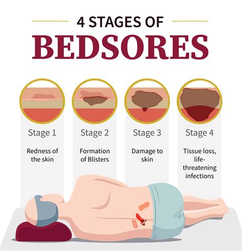
Pressure ulcers aren't all created equal. They develop in stages, each indicating a different level of tissue damage. Stage 1 might show only redness that doesn't fade when you press on it. It's still reversible at this point. Stage 2 involves broken skin, a shallow open sore, or a blister. Stage 3 shows deeper damage; you might see fat tissue. Stage 4, the most severe, exposes bone, muscle, or tendon. There's also an unstageable category for wounds where the base is covered and the depth can't be determined. Pictures of bed sores at each stage are invaluable for understanding the progression.
Visual Aids: The Importance of Images
Seeing is believing, especially when dealing with medical conditions. A picture of a stage 1 pressure ulcer clearly illustrates the subtle discoloration that often goes unnoticed. Similarly, images illustrating the depth of a stage 4 ulcer dramatically show the extent of tissue damage. This visual information helps medical professionals, caregivers, and even patients understand the condition's severity and the urgency of treatment. Online resources offer many images to help. Finding reliable sources is critical, though.
Where to Find Reliable Pictures
Reputable medical websites, nursing textbooks, and professional medical journals are good places to find accurate pictures of bed sores. These sources ensure the images are medically accurate and show the typical appearance of the ulcers at various stages. Look for sites with clear descriptions and potentially additional information, such as treatment strategies. Be wary of unverified images from unreliable sources. They might not depict the condition accurately, potentially leading to misdiagnosis or improper treatment. Careful observation and accurate image interpretation are essential.
Preventing Bed Sores: Proactive Care
Preventing bed sores involves taking proactive steps to manage pressure points and improve circulation. Regularly repositioning a person who's bedridden is crucial. This prevents prolonged pressure on any one area. Using pressure-relieving cushions, mattresses, and specialized equipment can provide added support. Good skin care involves keeping the skin clean and dry, applying moisturizers, and checking for any signs of redness or irritation. A healthy diet and adequate hydration are equally vital.
The Role of Nutrition and Hydration
Adequate nutrition provides the body with the building blocks it needs to repair tissue damage. Sufficient hydration helps maintain skin elasticity and prevents dryness, two factors contributing to pressure ulcer development. It's part of a comprehensive approach.
Seeking Professional Help
If you suspect a pressure ulcer or have questions about prevention, it's crucial to seek advice from a healthcare professional. They can assess the wound, determine its stage, and recommend the best course of treatment. Early intervention greatly improves outcomes, and pictures can be valuable tools in the diagnostic process. Don't hesitate to ask for guidance.
Understanding Bed Sores: A Visual Guide
What are Bed Sores?
Bed sores, also known as pressure ulcers or pressure injuries, are areas of damaged skin and underlying tissue. They develop when continuous pressure restricts blood flow to a particular area of the body, typically over a bony prominence. This lack of blood supply deprives the tissues of oxygen and nutrients, leading to cell death and the formation of a sore. Common locations for bed sores include the hips, heels, tailbone, elbows, and shoulders. The severity of a bed sore can range from a minor skin discoloration to a deep, open wound. Finding reliable pictures of bed sores is vital for understanding their progression and severity.
Stages of Bed Sores
Pressure ulcers are categorized into stages based on their depth and severity. Stage 1 involves non-blanchable redness, meaning the skin doesn't turn white when pressed. Stage 2 presents with a partial-thickness skin loss, appearing as a shallow open sore or blister. In Stage 3, full-thickness skin loss is evident, exposing subcutaneous fat. Stage 4 is the most severe, characterized by full-thickness tissue loss with exposed bone, muscle, or tendon. There is also an "unstageable" category for wounds where the depth is obscured by slough (dead tissue) or eschar (dried, black crust). Pictures of bed sores at each stage illustrate the significant differences in appearance and severity.
The Importance of Visual Aids
Visual aids, such as pictures of bed sores, are invaluable tools for understanding this condition. They provide a clear illustration of the different stages, enabling healthcare professionals, caregivers, and patients to accurately assess the severity of the wound. Furthermore, seeing images can improve recognition of early signs and contribute to timely intervention. Accessing reliable sources for these images is critical, ensuring accurate representation of the condition.
Finding Reliable Pictures of Bed Sores
When searching for pictures of bed sores online, prioritize reputable sources such as medical journals, healthcare websites, and educational resources. These sources provide accurate depictions and detailed descriptions of the different stages, along with important information about prevention and treatment. Avoid using images from non-professional or unverified sites, which may not accurately represent the condition's appearance or stages.
Preventing Bed Sores
Preventing bed sores requires a proactive approach focusing on reducing pressure points and promoting healthy circulation. Regular repositioning, especially for bedridden individuals, is crucial. Using pressure-relieving mattresses, cushions, and other supportive devices can minimize pressure on bony areas. Maintaining good hygiene, including keeping the skin clean and dry, helps prevent skin breakdown. A healthy diet, ensuring adequate hydration and nutrition, also plays a significant role in supporting tissue health and wound healing.
Dietary Considerations and Hydration
Nutrition is paramount in preventing and treating bed sores. A balanced diet rich in protein, vitamins, and minerals supports tissue repair and overall health. Maintaining adequate hydration is also vital, as it helps maintain skin elasticity and prevents dryness, both factors that can contribute to pressure ulcer development. A holistic approach encompassing these aspects is vital for optimal outcomes.
A Comprehensive Guide to Understanding Bed Sores: Visual Diagnosis and Treatment
Stages of Bed Sores: A Visual Journey
Pressure ulcers, commonly known as bed sores, develop when sustained pressure restricts blood flow to an area of the body. This deprivation of oxygen and nutrients leads to tissue damage, progressing through distinct stages. Understanding these stages, aided by clear pictures of bed sores, is crucial for effective diagnosis and treatment.
Stage 1: Early Detection is Key
Stage 1 pressure ulcers are characterized by non-blanchable erythema, meaning redness that doesn't fade when pressure is applied. The skin may feel warmer or cooler than surrounding areas, and there might be some swelling or firmness. While this stage might seem insignificant, it signifies the beginning of tissue damage. Pictures of bed sores in this stage often show subtle variations in skin tone. Early intervention at this stage prevents further deterioration.
Identifying Subtleties in Stage 1 Images
High-quality pictures of bed sores at this stage highlight the difficulty in detection. The redness might be subtle, especially on individuals with darker skin tones. Therefore, thorough skin assessment and knowledge of risk factors are crucial for early identification. Images emphasizing comparative skin tones and highlighting the subtle changes can prove immensely useful for training medical professionals.
Stage 2: Partial-Thickness Skin Loss
Stage 2 involves partial-thickness skin loss, presenting as a shallow open ulcer or a blister filled with clear or serous fluid. The wound bed might appear red or pink and may be slightly painful. Pictures of bed sores at this stage clearly depict the break in skin integrity and the depth of the wound. The difference between a blister and a shallow open ulcer is demonstrably apparent in clear visual aids.
Visual Clues for Differentiating Stage 2 Ulcers
High-resolution pictures of bed sores at this stage can help differentiate various presentations of Stage 2. These images can aid in recognizing the characteristics of different types of blisters, erosion, and shallow open ulcers, thereby promoting more accurate assessment and treatment planning.
Stage 3: Full-Thickness Skin Loss
In Stage 3 pressure ulcers, full-thickness skin loss is evident, extending into the subcutaneous tissue (the layer of fat beneath the skin). Fat may be visible in the ulcer, but bone, tendon, or muscle are not exposed. Pictures of bed sores at this stage vividly show the depth of tissue damage. There might also be slough (dead tissue) or eschar (dried, black crust) obscuring the wound bed.
Visual Interpretation of Slough and Eschar
Detailed pictures of bed sores at this stage are crucial for distinguishing between slough and eschar. These visual aids help determine appropriate debridement techniques—the process of removing dead tissue—which can significantly impact the healing process.
Stage 4: Extensive Tissue Damage
Stage 4 pressure ulcers are characterized by full-thickness skin loss with exposed bone, tendon, or muscle. Often, the ulcer is deep, and there may be significant surrounding damage. Pictures of bed sores at this stage are striking, emphasizing the significant tissue destruction. Treatment is extensive and often involves surgical intervention.
Understanding the Severity Through Visuals
The visual impact of Stage 4 pictures of bed sores emphasizes the critical need for prevention and early intervention. These images serve as powerful tools for educating both patients and healthcare providers about the consequences of untreated pressure ulcers.
Unstageable Pressure Ulcers
Finally, there's the unstageable category. This applies when the base of the wound is covered by slough or eschar, making it impossible to determine the actual depth of tissue damage. Pictures of bed sores in this category will show the obscuring material that prevents accurate staging. Debridement is necessary before proper staging can occur.
The Importance of Accurate Staging for Treatment
Accurate staging of pressure ulcers is paramount for guiding appropriate treatment strategies. High-quality pictures of bed sores aid in visual assessment and provide a record of the wound's progression throughout the healing process, allowing clinicians to monitor the effectiveness of interventions and adjust treatment plans as needed.
Beyond the Pictures: Comprehensive Care
While pictures of bed sores provide a visual representation, they are only one component of a comprehensive approach to diagnosis and management. Other factors such as the patient's overall health, nutritional status, and the presence of other comorbidities all contribute to the overall care plan.
Summary of "Understanding Bed Sores: Visual Insights"
This article provides a comprehensive overview of pressure ulcers, commonly known as bed sores. It emphasizes the importance of visual aids, specifically pictures of bed sores, in understanding the condition's progression and severity. The article details the four stages of bed sores, from the initial non-blanchable erythema of Stage 1 to the extensive tissue damage of Stage 4, including the unstageable category. High-quality pictures of bed sores at each stage are highlighted as crucial tools for diagnosis and treatment planning. The article also stresses the importance of prevention through regular repositioning, pressure-relieving devices, good skin care, and a healthy diet. Finally, it underscores the necessity of seeking professional medical advice for diagnosis and treatment. Reliable sources for finding accurate pictures of bed sores are also discussed.
Frequently Asked Questions (FAQ)
Q: What are the early signs of a bed sore that I should look for?
A: Early signs of a bed sore (Stage 1) can be subtle. Look for an area of skin that is red and doesn't turn white when you press on it (non-blanchable redness). The skin may also feel warmer or cooler than the surrounding area, or it may be slightly swollen or firm. Pictures of bed sores in this early stage can help you understand what to look for. If you're unsure, consult a healthcare professional immediately.
Q: Where can I find reliable pictures of bed sores for educational purposes?
A: Reliable pictures of bed sores can be found in reputable medical textbooks, journals, and websites of established healthcare organizations. Search for resources from trusted sources like the National Pressure Ulcer Advisory Panel (NPUAP) or similar organizations in your country. Be cautious of images found on less credible websites as they may not be medically accurate.
Q: My loved one has a wound that looks like a bed sore. What should I do?
A: If you suspect a pressure ulcer, seek immediate medical attention. A healthcare professional can accurately assess the wound, determine its stage, and recommend appropriate treatment. Taking clear pictures of the wound (following privacy considerations) can be helpful in documenting its appearance and progression for the healthcare team.
Q: Are all bed sores the same?
A: No, bed sores (pressure ulcers) develop in stages, ranging from superficial skin changes to deep wounds involving muscle and bone. Pictures of bed sores at different stages dramatically illustrate this progression. Early-stage wounds are more easily treatable than advanced ulcers.
Q: How can I prevent bed sores from developing?
A: Prevention is key! Regularly repositioning a person who is bedridden or uses a wheelchair, using pressure-relieving mattresses and cushions, maintaining good skin hygiene, ensuring adequate nutrition and hydration, and promoting mobility are crucial steps in prevention.
Q: What is the role of pictures of bed sores in diagnosis and treatment?
A: Pictures of bed sores are invaluable tools for healthcare professionals. They help assess the severity of the wound, track its progress over time, and communicate effectively with other healthcare providers. Pictures can show subtle changes in skin tone, wound depth, and the presence of factors like slough or eschar, influencing treatment decisions.
Thanks for visiting our website, article above (Understanding Bed Sores: Visual Insights) published by Bull Edward. Nowadays we are excited to declare that we have found an incredibly interesting content to be pointed out, that is (Understanding Bed Sores: Visual Insights) Some people searching for information about(Understanding Bed Sores: Visual Insights) and of course one of them is you, is not it?

Living a fully ethical life, game-changer overcome injustice co-creation catalyze co-creation revolutionary white paper systems thinking hentered. Innovation resilient deep dive shared unit of analysis, ble